The moment you see a face, your brain sends a small electrical signal. Imagine a high school student recording that signal using her own laptop. Brain mapping for students makes this possible. Affordable EEG headsets and free software turn raw brain signals into colorful maps of attention, memory, and sleep. As a result, classrooms can become small neuroscience labs, and curious teenagers can do real research. However, this power comes with responsibility. Students must understand the technology, receive ethics training, and be supervised by adults. This guide explains the tools, methods, and safety rules. It also offers beginner-friendly project ideas. Let’s start with the basics.
What Is Brain Mapping? (Simple Definition)
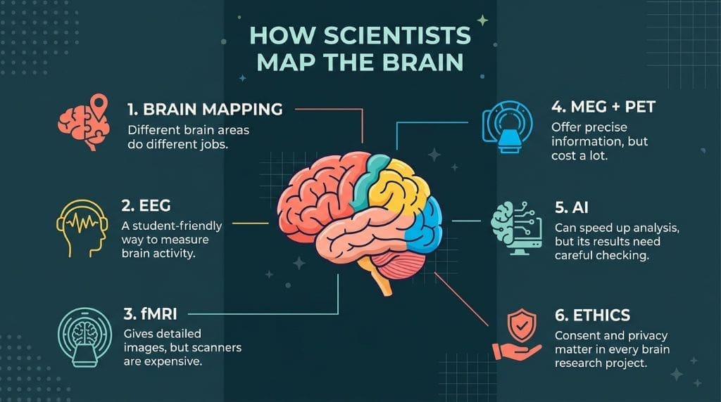
Brain mapping shows the brain regions responsible for specific functions. To do this, scientists observe blood flow, electrical activity, and magnetic fields. For instance, neurons fire as you read this sentence; the signals they produce cause small changes detectable by scanners. Researchers therefore produce maps of active areas. It should be noted that these maps are not photographs of thoughts; rather, they display statistical patterns derived from data collected from many people. The way brain mapping works varies depending on the technology used.

In reality, each of the tools captures a different type of signal: EEG measures electrical activity via electrodes placed on the scalp, fMRI monitors changes in blood oxygenation, MEG detects the magnetic fields produced by neurons, and PET employs radioactive tracers to show metabolism. Students can begin by working with free datasets. Taking this approach reduces costs and ensures safety, which in turn helps them build confidence before conducting actual recordings.
Brain Imaging Technologies Compared
It is difficult for people just starting to choose a method, since each technique sacrifices some detail to be either faster or cheaper. Spatial resolution refers to a scanner’s ability to show where activity is occurring, and temporal resolution refers to how quickly it can monitor changes. For instance, fMRI does a good job of showing the locations but is slow. EEG is fast but has difficulty with signals from deep in the brain. MEG is fast and has better spatial accuracy.
On the contrary, MEG machines cost millions of dollars. PET provides information on brain chemistry but requires radioactive tracers. For students, it is just as important to have access to the equipment as it is for it to function well, which is why EEG is generally the best option to begin with. The hardware is inexpensive, and the software is available at no cost. The table below compares the resolution, cost, and use cases, and rates each tool’s accessibility for students. You should select tools that match your project objectives.
fMRI — How It Works, Pros & Cons
Functional MRI (fMRI) can detect oxygenated blood in active brain regions. Since neurons require energy to communicate, blood flow increases to meet this demand, bringing in fresh oxygen and leading to the blood-oxygen-level-dependent (BOLD) signal, which forms the basis of most fMRI studies (He & Liu, 2008). For instance, performing a memory task can increase activity in the hippocampus. The scanner splits the brain into small cubes, known as voxels, each only a few millimeters wide, so that the maps can show exact locations.


Yet fMRI has obvious limitations. Since the BOLD signal peaks several seconds after neurons fire, rapid events tend to blend. Moreover, participants must remain still inside the noisy scanner, and even minor movements can cause image distortion. Scanner time can also be costly. To sum up, although fMRI provides good spatial detail and extensive coverage of the brain, it is accompanied by high cost, noise, sensitivity to motion, and a delayed signal (Warbrick, 2022).
Given these difficulties, students generally prefer to use open fMRI datasets rather than conduct their own scans. Instead of having to buy or book time on a scanner, they can work with the actual brain data from previously shared recordings.
EEG — Electrical Brain Activity
EEG records electrical activity using electrodes placed on the scalp; the resulting patterns show millions of neurons firing simultaneously. The technique can record timing to within milliseconds. For instance, the N170 wave appears when a face is viewed. Students frequently carry out this experiment. Headsets currently cost a few hundred dollars, and free software such as EEGLAB is available for analysis. For these reasons, EEG is the most appropriate technique for classroom budgets.
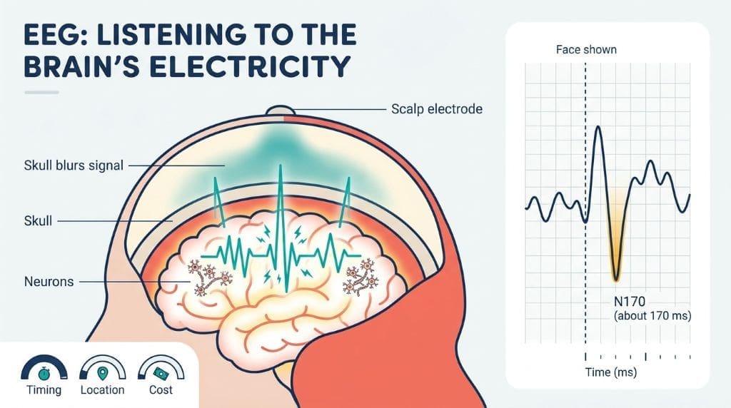
EEG has poor spatial accuracy because the skull spreads the electrical signals, and recordings are influenced by muscle tension and eye blinks. Students are instructed to eliminate these artifacts right from the start. As long as they are properly trained, the recordings stay clean and safe. It is the most accessible brain imaging technique for students, provided they have patience, can carry out some coding, and design their experiments carefully. The electrode caps must be used with conductive gel and handled with care. The setup generally takes new learners about thirty minutes, which helps them build up good laboratory habits.
MEG & PET — Advanced Techniques
MEG measures magnetic fields generated by neural currents. Its sensors require extreme cooling and shielded rooms. Still, it tracks brain activity in milliseconds. Unlike EEG signals, magnetic signals pass cleanly through the skull. As a result, MEG localizes sources more accurately. For example, clinicians use MEG before epilepsy surgery. PET works differently by tracking radioactive tracers. Those tracers reveal metabolism, receptors, and blood flow. Because of the radiation, PET is not used with healthy teenagers in school projects. Each scan also costs a lot, so both tools stay research-grade for most classrooms.
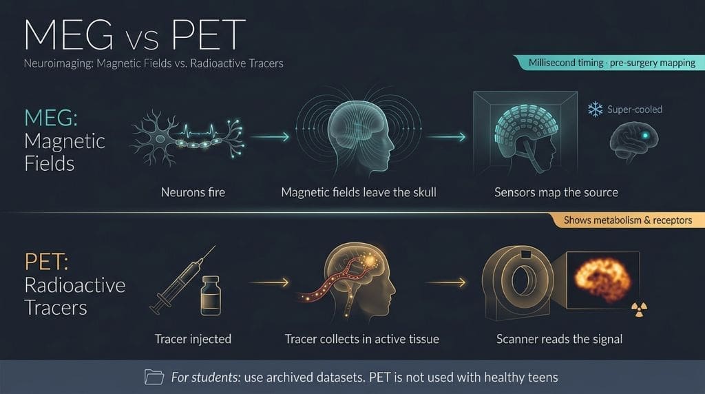

It is difficult to access MEG and PET equipment, but students can still gain knowledge by studying archived recordings available online. It is useful to use these datasets in class because both techniques require specialized training and there are only a few places with MEG systems.
Comparison Table: Resolution, Cost, Use Case, and Student Accessibility
The table given below illustrates the trade-offs involved with the four methods. The resolution figures are taken from published reviews of neuroimaging studies (Yen et al., 2023), and the costs are only approximate, so the figures provided by your school might differ. The accessibility ratings take into account the required equipment, training needs, and budgetary considerations. EEG has the highest rating since it is cheap. fMRI has a lower rating because it requires specialized facilities and strict movement restrictions. MEG and PET are the least accessible for use in classrooms.
| Method | Spatial Resolution | Temporal Resolution | Typical Cost | Best Use Case | Student Accessibility |
| EEG | Low (centimeters) | Excellent (milliseconds) | Low | Timing studies, sleep, ERPs | High: affordable headsets, free software |
| fMRI | High (millimeters) | Slow (seconds) | High | Localization, connectivity | Medium: shared or open data |
| MEG | Medium-high (millimeters to about 1 cm) | Excellent (milliseconds) | Very high | Epilepsy, fast dynamics | Low: mostly shared datasets |
| PET | Medium | Slow | Very high | Metabolism, receptors | Low: mostly shared datasets |
You aren’t limited to using equipment as your only resource, since some universities allow students to use their own scanners, and public datasets let you get some practice without needing special hardware. Most importantly, choose tools appropriate for your research question; a simple event-related potential (ERP) study is better than one so complex you won’t be able to finish it. Begin with projects that you can handle and then move on to more challenging ones as your knowledge and skills increase.
How AI Is Transforming Brain Mapping Research
Currently, AI is capable of carrying out jobs which previously had taken weeks. For instance, algorithms can automatically identify different areas of the brain, and machine learning is able to predict brain age based on scans. AI tools can also highlight mild abnormalities for doctors to examine. Moreover, the use of AI-assisted EEG interpretation is being made to help with epilepsy care (Singh et al., 2022). Since these tools learn patterns from thousands of examples, the analyses have become quicker and more consistent.
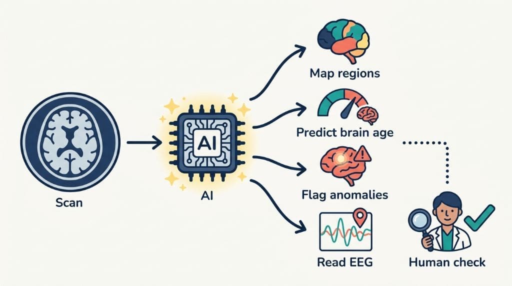
Since the training data is biased, the results will also be biased, and a model created in one hospital may not function in another, so it is important to verify the results at various sites. Students can take part in this by comparing the model’s predictions with those made by experts. Harmonization tools help reduce differences between scanners, but it is still essential to maintain transparency to ensure trustworthy findings. Humans should review each automated output. Although AI accelerates the discovery process, it cannot replace careful thought. Using these tools helps develop the skills needed for careers in data-driven neuroscience, and nowadays, courses teach coding alongside biology.
Brain Mapping for Students: Safe Project Ideas to Start With
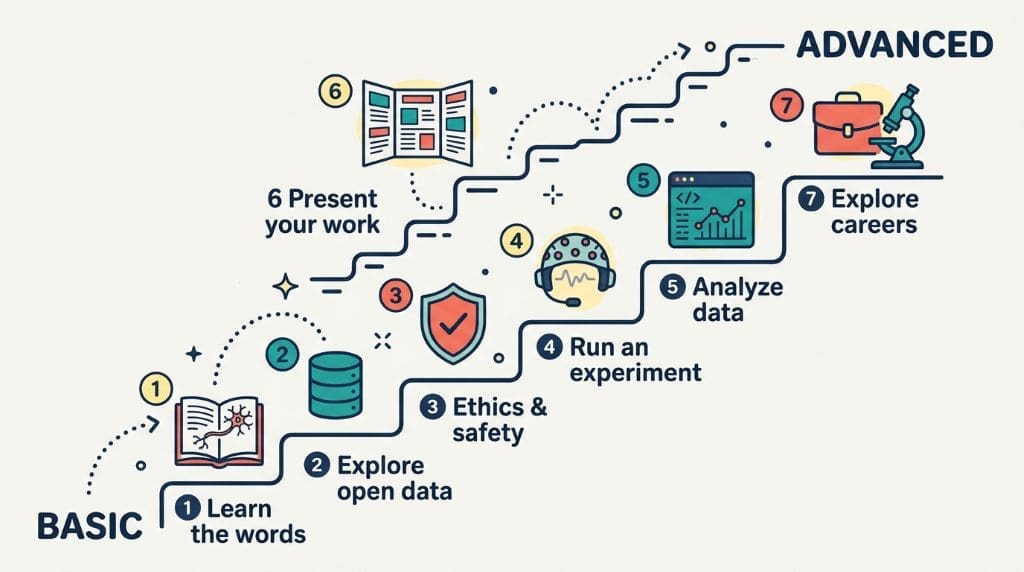
Good projects start by asking simple, testable questions. Safety, consent, and supervision must always be given the highest priority. The list mentioned below progresses from basic skills to more advanced ones. New learners should begin by finding out how to eliminate unwanted signals from data. Each stage is based on the one that came before it, and teachers can modify the difficulty level according to each student’s ability. In this way, all students can proceed safely without taking any risks. Most projects make use of EEG data or data from public sources. Whenever a project involves human subjects, it has to receive approval from an adult mentor. Students ought to begin with archived recordings in order to keep the volunteers safe and to obtain better data.
Getting started with live recordings too early may result in errors. Therefore, adhere to these seven steps, progressing from the basic to the more advanced ones. Each step has a specific learning objective. Feedback from mentors helps to keep you on the right path, and achieving early successes gives you the confidence needed for more difficult projects.
Case Studies: Real Student Brain Mapping Research
The course reports illustrate the abilities of undergraduate students. For instance, Nyhus and Curtis (2016) outlined a project involving an ERP study over a period of one semester in which the students designed their own EEG experiments, gathered the data, eliminated the unwanted signals, and carried out statistical analysis. Since some of the students found statistics and programming difficult, the instructors provided additional support and held tutorials. The report recommends making use of public datasets and free tools, a method which is the one adopted in the project ideas mentioned above.
However, access to equipment remains a barrier. Some universities share scanner time with local schools, and many summer programs offer intensive lab experience. Student conferences also give newcomers a chance to present early work. These events encourage careful methods and honest reporting. For example, poster sessions often welcome first-time presenters, and feedback from judges helps improve both analysis and communication.
Reproducibility is what makes science reliable. Poldrack and colleagues (2017) pointed out common problems in neuroimaging. Small sample sizes can lead to unreliable brain maps, and flexible analytical methods can lead researchers to find patterns that might not be real. For example, a single dataset could support many different claims. Errors from making too many comparisons can also lead to false positives. That’s why students should preregister their analysis plans and use open data and shared code so others can check their work. These steps may seem slow, but they help build trust.
The classroom posters should also be held to the same standards. Since teachers can catch many errors early on, it is important that the methods section be written carefully. Performing a power analysis will help prevent wasted effort, and using statistical corrections ensures that the results remain honest. These practices are useful for students in any subject, not just those studying neuroscience.
Research Ethics Guidelines for Students
Ethics plays a role in safeguarding volunteers, the data, and public confidence in brain mapping projects involving students. Shen and his team have pointed out seven issues that remain unresolved in the context of portable neuroimaging. Among these, informed consent is the most important aspect of field research; participants must be made aware of the risks, benefits, and how their data will be used. It is necessary to ensure privacy and security through encryption and access restrictions. Since the data is stored in the cloud, brain data can be sent to any location. There are difficult questions involved in the case of returning incidental findings. For example, who should explain an unexpected anomaly to a teenager? Algorithmic bias may negatively affect underrepresented communities. Moreover, participants should have access to their own scans. Consequently, schools should have clear, written protocols; for example, review boards guide research involving human subjects. Students must complete ethics training before collecting any data. Of course, teacher supervision should continue right throughout the project.
To sum up, simple checklists can avoid serious errors. If there is any uncertainty, one should first ask a mentor. Each decision must put the volunteer first rather than the results.
Real clinical data is usually unnecessary for classroom projects; it is better to use tutorial datasets. For instance, EEGLAB provides sample recordings for practical use. Storage archives that omit personal details help to protect privacy. Students should nevertheless be taught how to obtain consent. Role-playing is a simple method of practicing these skills, and having guest speakers who are members of the review board makes the experience realistic. When students publish their work, they must report the data honestly because fake or altered data undermines credibility. Supervisors should check all final analyses. Ethics should be covered in each lab meeting. That is why we provide consent training at the beginning of each semester. This, in turn, helps students become ethical researchers in the long term. Safety checks are important for both students and participants. Screening forms can identify conditions such as epilepsy before the sessions take place. Looking after the electrodes helps to avoid skin problems. Taking breaks and drinking water keeps everyone comfortable. Thorough cleaning is also important for good science.
How This Brain Mapping Student Research Guide Was Compiled
The guide draws on practical classroom experience and evidence reviewed by peers. The technical sections refer to the open-access reviews listed below; for instance, the ones concerning EEG, fMRI, and MEG are based on published research. The advice regarding ethics comes from Shen and his colleagues, and the tips on reproducibility are from Poldrack and his colleagues. The guide contains no private data or unverified statements.
Frequently Asked Questions
EEG is easiest for classrooms. Headsets are affordable, and software is free. Students see results within one session.
Yes, with supervision. Many labs welcome students for summer projects. Public datasets also offer genuine research experience.
Yes, with parental permission and adult supervision. EEG records signals without radiation or injections. Proper electrode care prevents skin irritation.
A basic EEG setup costs a few hundred dollars. Free toolboxes handle all analysis. Public datasets cost nothing at all.
Open repositories and tutorial archives share real recordings. EEGLAB includes practice datasets for beginners. Teachers can download them instantly.
Teachers should complete safety and ethics training first. University outreach programs offer short courses. Mentors can co-supervise early projects.
References
- Delorme, A., & Makeig, S. (2004). EEGLAB: An open source toolbox for analysis of single-trial EEG dynamics including independent component analysis. Journal of Neuroscience Methods, 134(1), 9–21. https://doi.org/10.1016/j.jneumeth.2003.10.009
- Fred, A. L., Kumar, S. N., Haridhas, A. K., Ghosh, S., Bhuvana, H. P., Sim, W. K. J., Vimalan, V., Givo, F. A. S., Jousmäki, V., Padmanabhan, P., & Gulyás, B. (2022). A brief introduction to magnetoencephalography (MEG) and its clinical applications. Brain Sciences, 12(6), 788. https://doi.org/10.3390/brainsci12060788
- He, B., & Liu, Z. (2008). Multimodal functional neuroimaging: Integrating functional MRI and EEG/MEG. IEEE Reviews in Biomedical Engineering, 1, 23–40. https://doi.org/10.1109/RBME.2008.2008233
- Nyhus, E., & Curtis, N. (2016). Incorporating an ERP Project into Undergraduate Instruction. PubMed, 14(2), A91-6. https://pubmed.ncbi.nlm.nih.gov/27385925
- Poldrack, R. A., Baker, C. I., Durnez, J., Gorgolewski, K. J., Matthews, P. M., Munafò, M. R., Nichols, T. E., Poline, J.-B., Vul, E., & Yarkoni, T. (2017). Scanning the horizon: Towards transparent and reproducible neuroimaging research. Nature Reviews Neuroscience, 18(2), 115–126. https://doi.org/10.1038/nrn.2016.167
- Shen, F. X., Wolf, S. M., Gonzalez, R. G., & Garwood, M. (2020). Ethical issues posed by field research using highly portable and cloud-enabled neuroimaging. Neuron, 105(5), 771–775. https://doi.org/10.1016/j.neuron.2020.01.041
- Singh, N. M., Harrod, J. B., Subramanian, S., Robinson, M., Chang, K., Cetin-Karayumak, S., Dalca, A. V., Eickhoff, S., Fox, M., Franke, L., Golland, P., Haehn, D., Iglesias, J. E., O’Donnell, L. J., Ou, Y., Rathi, Y., Siddiqi, S. H., Sun, H., Westover, M. B., Whitfield-Gabrieli, S., & Gollub, R. L. (2022). How machine learning is powering neuroimaging to improve brain health. Neuroinformatics, 20(4), 1093–1109. https://doi.org/10.1007/s12021-022-09572-9
- Warbrick, T. (2022). Simultaneous EEG-fMRI: What have we learned and what does the future hold? Sensors, 22(6), 2262. https://doi.org/10.3390/s22062262
- Yen, C., Lin, C.-L., & Chiang, M.-C. (2023). Exploring the frontiers of neuroimaging: A review of recent advances in understanding brain functioning and disorders. Life, 13(7), 1472. https://doi.org/10.3390/life13071472